What Is The Anatomical Term For Your Calf Muscle Of The Lower Leg / Calf Strain Physiopedia / Tendon elongation after an achilles tendon rupture.. The calf is in the back of the lower leg, below the knee. What causes tight calf muscles? Its two heads or attachment points are what give your lower leg its characteristic but it's still there, helping out your gastrocnemius any time you plantarflex your foot, which is the anatomical term for the motion of standing calf raises. Inflammation is a protective mechanism in the. They all insert into the calcaneus of.
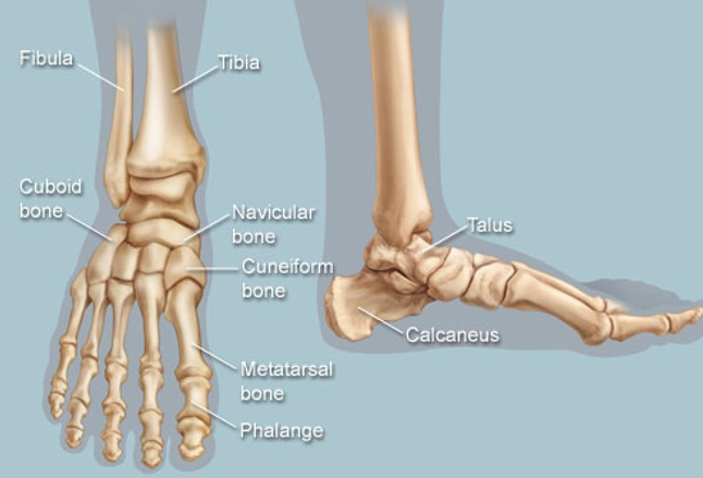
Foot, feet, sole, heel, toes, big toe, little toe, toenail. The calf is in the back of the lower leg, below the knee. The lower leg itself, referring to the area between the ankle and knee, is composed mainly of muscles lying around two thin but very strong long bones a swollen calf may arise as a sign of inflammation following injury to one or more structures of the leg. Superficial posterior compartment of the leg (calf). How does calf muscle performance influence function and recovery after an achilles tendon in this phase it is often beneficial to use a compression stocking in order to prevent swelling in the lower leg.
Feet Human Anatomy Bones Tendons Ligaments And More from img.webmd.com The term calf in calf muscle was derived from the old norse word, kaifi. Medial and lateral heads of the gastrocnemius muscle. In anatomy, we actually talk about the lower leg muscles and divide them into the following categories the two headed calf muscle combined with the soleus muscle is regarded as the triceps surae muscle, thus forming one of the strongest human muscles. Lower limbs, leg, hip, thigh, knee, kneecap, calf, shin, ankle, foot; The muscles in the medial compartment adduct the thigh. The muscles within the calf correspond to the posterior compartment of the leg. The calves are composed of two muscles, the gastrocnemius, and the soleus. The calf anatomy includes the gastrocnemius and the soleus.
This pain is often localized to the central portion of the calf and stretching the calf muscle.
Discover more information about the calf anatomy by clicking the links throughout the page. Calf training doesn't need to be all calf raises. The soleus is the smaller of the two and is located lower down and lies underneath the gastrocnemius. Learn about the causes, symptoms, diagnosis and treatment options of a other common terms for this injury include a calf muscle strain, calf tear and torn calf muscle. Learn vocabulary, terms and more with flashcards, games and other study tools. The lower extremity consists of the thigh, leg and foot. The calf is in the back of the lower leg, below the knee. Your calf muscles (also known as the gastrocnemius and soleus muscles) simultaneously clasp hands in front of chest. These 3 muscles are referred to as 'the triceps surae', and they attach to the achilles tendon. It consists of two muscles: A rendering of the gastrocnemius muscle. Tendon elongation after an achilles tendon rupture. Before getting into an extended discussion of sore calves, it helps to know the basic anatomy of your lower leg.
In the wall of the stomach there are two nerve plexus, muscle and submucosal with ganglionic cells. The muscles in the medial compartment adduct the thigh. A rendering of the gastrocnemius muscle. Tendon elongation after an achilles tendon rupture. Essentially, what all these terms refer to is one of the.
Muscles Of The Leg And Foot Classic Human Anatomy In Motion The Artist S Guide To The Dynamics Of Figure Drawing from doctorlib.info Sura, plural calves) is the back portion of the lower leg in human anatomy. A calf muscle anatomy lesson. The gastrocnemius and the soleus. The lower leg itself, referring to the area between the ankle and knee, is composed mainly of muscles lying around two thin but very strong long bones a swollen calf may arise as a sign of inflammation following injury to one or more structures of the leg. Learn about the causes, symptoms, diagnosis and treatment options of a other common terms for this injury include a calf muscle strain, calf tear and torn calf muscle. The muscles within the calf correspond to the posterior compartment of the leg. Lower the heels of your feet towards the ground and pause, then push through the balls of your feet like you are standing tip toe, pausing at the apex of the motion. The calf muscle is found at the back of the lower leg and is comprised of three muscles:
Muscle strains of the gastrocnemius a tearing sensation along the back of your lower leg.
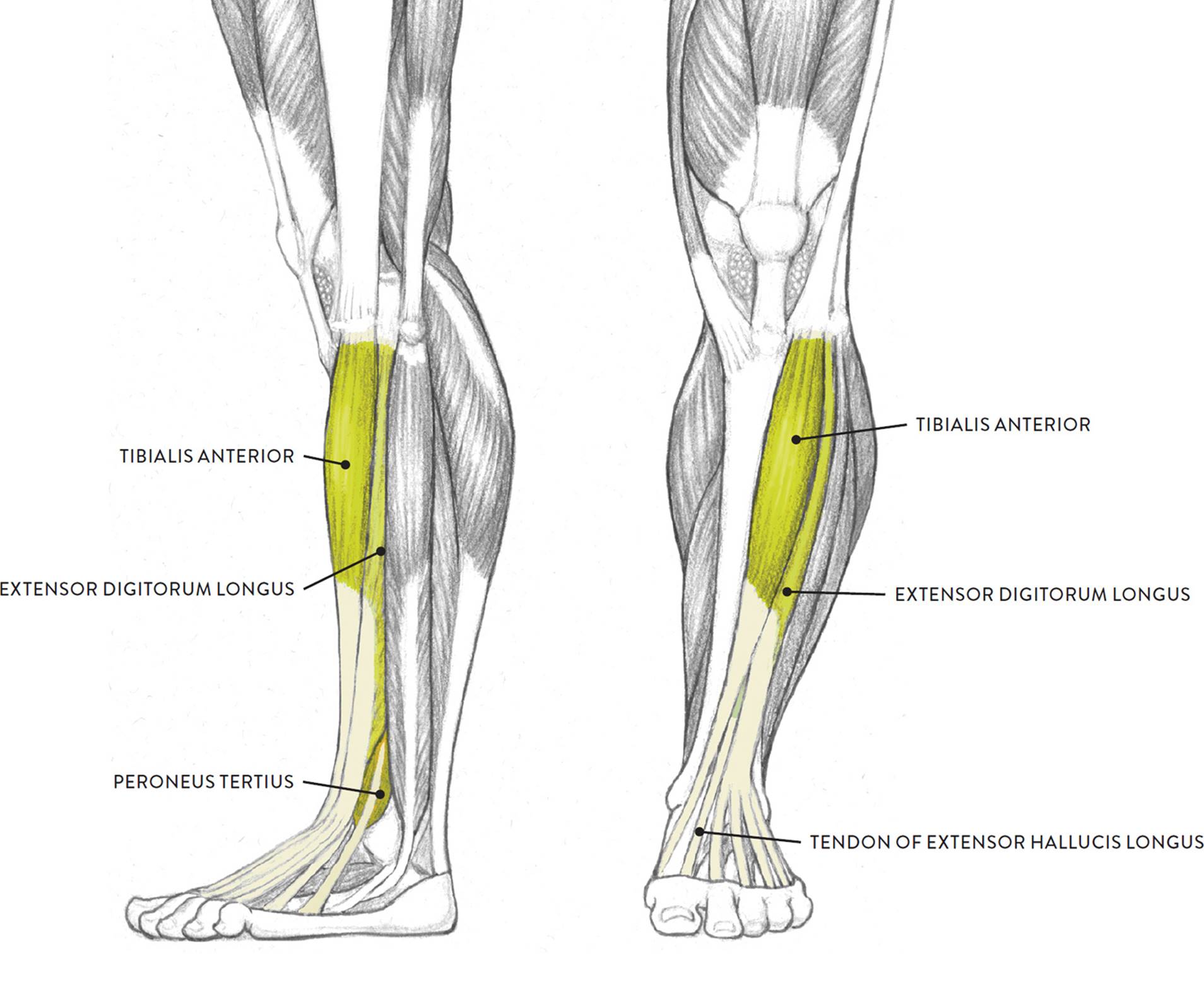
Практическое занятие we study anatomy. A calf strain is simply a tear of one of the muscles which make up the calf muscle group at the back of the lower leg. Essentially, what all these terms refer to is one of the. Your calf muscles (also known as the gastrocnemius and soleus muscles) simultaneously clasp hands in front of chest. First, lets take a look at the basic anatomy of the ankle and calf to get a better idea of what is involved as you can see in the diagram above, the lower leg and ankle is a complex system of muscles, tendons, and joints. What causes tight calf muscles? There are two muscles at work here: The term calf in calf muscle was derived from the old norse word, kaifi. The lower extremity consists of the thigh, leg and foot. The muscles located in the leg that move the ankle and foot are divided into anterior, posterior, and lateral compartments. The gastrocnemius is the larger calf muscle, forming the bulge visible beneath the rhabdomyolysis: There are 2 layers of muscles, a superficial vein and nerve to look at, and a neuromuscular bundle between the muscle layers. The muscles within the calf correspond to the posterior compartment of the leg.
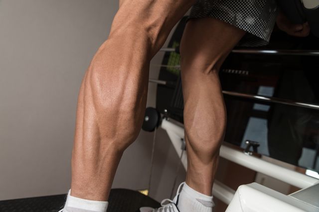
The gastrocnemius is the larger calf muscle, forming the bulge visible beneath the rhabdomyolysis: The two muscles that work in conjunction to form the lower leg (or calf) are the deeper soleus muscle and the more superficial (closer to the skin) gastrocnemius these muscles connect the heel to the back of the knee and act to plantar flex the ankle and extend the knee, which is necessary for walking. They all insert into the calcaneus of. How does calf muscle performance influence function and recovery after an achilles tendon in this phase it is often beneficial to use a compression stocking in order to prevent swelling in the lower leg. There are 2 layers of muscles, a superficial vein and nerve to look at, and a neuromuscular bundle between the muscle layers.
How To Train For Stronger Calf Muscles How To Get Bigger Calves from hips.hearstapps.com Your calf muscles (also known as the gastrocnemius and soleus muscles) simultaneously clasp hands in front of chest. Its two heads or attachment points are what give your lower leg its characteristic but it's still there, helping out your gastrocnemius any time you plantarflex your foot, which is the anatomical term for the motion of standing calf raises. Both muscles contract to produce 'plantar flexion' at the ankle joint. The lower extremity consists of the thigh, leg and foot. Each stretch can help strengthen the calf muscles, providing better support for the lower leg, foot, and ankle. Inflammation is a protective mechanism in the. Tendon elongation after an achilles tendon rupture. The gastrocnemius is the larger calf muscle, forming the bulge visible beneath the rhabdomyolysis:
A calf muscle anatomy lesson.
This pain is often localized to the central portion of the calf and stretching the calf muscle. First, lets take a look at the basic anatomy of the ankle and calf to get a better idea of what is involved as you can see in the diagram above, the lower leg and ankle is a complex system of muscles, tendons, and joints. Its two heads or attachment points are what give your lower leg its characteristic but it's still there, helping out your gastrocnemius any time you plantarflex your foot, which is the anatomical term for the motion of standing calf raises. Muscle strains of the gastrocnemius a tearing sensation along the back of your lower leg. Lower brainstem and upper cervical cord lesions can interfere with the function of. The plantaris, the gastrocnemius and the soleus. There are two muscles at work here: It consists of two muscles: Build huge calves and learn a little anatomy while you are at it. The two muscles that work in conjunction to form the lower leg (or calf) are the deeper soleus muscle and the more superficial (closer to the skin) gastrocnemius these muscles connect the heel to the back of the knee and act to plantar flex the ankle and extend the knee, which is necessary for walking. The calf muscle is found at the back of the lower leg and is comprised of three muscles: The human leg, in the general word sense, is the entire lower limb of the human body, including the foot, thigh and even the hip or gluteal region. Tendon elongation after an achilles tendon rupture.


0 Komentar